Share
Acute angle closure causes a sudden very high pressure. Symptoms can be severe, and patients usually seek help from a doctor or optometrist. If the condition is promptly recognised and appropriate referrals made, urgent treatment can avoid permanent damage.
Less common than Open angle glaucoma, Angle closure can be far more serious. Angle closure can be:
- Acute: sudden onset of very high eye pressure. Symptoms of pain, blurred vision, a
 nd seeing haloes around lights.
 - Intermittent, or
- Chronic: pressure builds up slowly (silently), as with primary open angle glaucoma, but can be more aggressive.
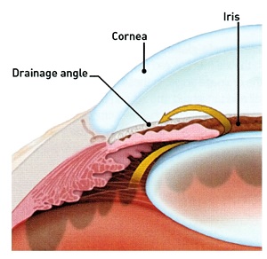
The ‘angle’ of the eye is the space between the iris (coloured part) and cornea (clear outer part at the front). It is important for eye pressure because the trabecular meshwork (where fluid drains out of the eye) is in the angle.
If the angle closes or even just narrows, fluid can build up causing elevated eye pressure. Elevated eye pressure damages the optic nerve. Glaucoma is optic nerve damage.
Detection
Acute angle closure causes a sudden very high pressure. Symptoms can be severe, and patients usually seek help from a doctor or optometrist. If the condition is promptly recognised and appropriate referrals made, urgent treatment can avoid permanent damage.
Chronic angle closure is usually asymptomatic. Elevated eye pressure can be detected by IOP measurement by an Optometrist or Ophthalmologist. All patients with an elevated eye pressure should have gonioscopy to make sure they do not have angle closure. Gonioscopy is where a special lens is used to view the angle to see if it is open or closed. It is an important part of the eye examination to assess patients who may have glaucoma.
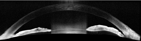
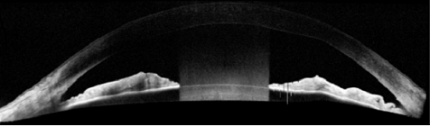
Anterior segment OCT is a scan that can image the angle structures. This can help illustrate if the angle is narrow, and document changes after interventions.
Image: Anterior segment OCT images of a patient with angle closure, before and after laser iridotomy.
UBM
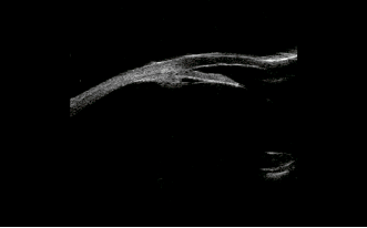
UBM is a special type of ultrasound which can give detail of angle structures, particularly useful in special or unusual cases.
As for patients with all types of glaucoma, visual field assessment and optic disc imaging are important for assessing the degree of damage to the optic nerve which has occurred from the high pressure.
Image: UBM of a patient with ‘plateau iris syndrome’, a less common type of angle closure glaucoma.
Surgical Interventions
Laser peripheral iridotomy (LPI) is the most important.
- This procedure can be performed in office. A small hole is made at the outer edge of the iris which helps open the angle.
- LPI can lower the pressure, and prevent angle closure getting worse.
- In patients with an asymptomatic narrow angle LPI can prevent acute attacks
Laser iridoplasty (less commonly performed)
- A different type of laser is used to flatten the peripheral iris, draw it out from the angle.
- Sometimes used in special situations where LPI is not enough (such as plateau iris).
Lens extraction/IOL (cataract surgery) can be extremely useful for patients with angle closure, especially if there is also cataract present.
When people have cataract surgery the lens of the eye is removed and replaced with an artificial lens (IOL) which is thinner than the original. Because the lens sits just behind the iris, when the new thinner lens is in place the drainage angle opens up.
- Many cases of angle closure occur because the lens has become thicker and is starting to form a cataract.
- Cataract surgery is a commonly performed surgical procedure with very high success rate, although can be challenging in eyes with angle closure
Trabeculectomy (glaucoma filtering surgery)
- A small hole is made in the top of the eye to allow fluid to drain into a bubble under the skin of the eye. It provides an alternative route for fluid to leave the eye.
- Very useful in angle closure where other treatments have failed to control the pressure.
- Also where a very low pressure is required to protect eyes with advanced glaucoma.
During the past decade, glaucoma care in the UK has changed dramatically, with more non-medical ophthalmic practitioners involved. As such, optometrists and other non-medical professionals have been delivering laser treatments including SLT in the Hospital Eye Service (HES).
Sustained drug delivery via an implant could reduce the reliance on daily eye drops and ensure that the correct amount of medicine is delivered consistently, every day.
With the rising numbers of patients diagnosed with glaucoma – and oversubscription of public ophthalmology departments – the Glaucoma Community Collaborative Care Program at the Royal Victorian Eye and Ear Hospital in Melbourne is designed to alleviate some of this pressure.