There are many different kinds of laser used in ophthalmology and specifically in glaucoma. A laser is amplified light energy which can be targeted to different parts of the eye with minimal or no damage to surrounding structures.
Different lasers are used to treat open and closed angle glaucoma. Laser can be applied to the iris or the trabecular meshwork to allow aqueous fluid to flow more effectively within the eye, and drain better by the normal drainage channels within the eye.
Selective Laser Trabeculoplasty (SLT)
What is it?
SLT is a laser procedure used to treat glaucoma and reduce eye (intraocular) pressure by improving fluid outflow through drainage pores located within the trabecular meshwork.
How does it work?
SLT uses short pulses of low-energy light to improve drainage of aqueous fluid in order to lower eye pressure.
In suitable patients it may be used to try and lower the eye pressure without the need for ongoing drops, or as a supplement to existing medications. The aim of the laser treatment is to lower the eye pressure and it does not improve vision. There is a very low complication rate.
Who is it suitable for?
Your eye specialist (ophthalmologist) may recommend SLT as initial treatment:
- If your eye pressure is not adequately controlled with current medication(s)
- If your glaucoma is worsening
- If you are experiencing problems with your glaucoma medication(s)
What are the benefits?
SLT is considered a safe and effective procedure with few risks. SLT lowers the eye pressure by an average of 25% in 74%-85% of patients treated. The duration of the effect of the laser is variable, with an average benefit of around 3-4 years. One benefit of SLT is that it may be repeated but success with repeat treatments is lower each time.
It may be necessary to supplement SLT with ongoing glaucoma medication. For those who do not respond to SLT, your doctor will discuss alternative treatment options.
Before the procedure
Please take all medication as normal or as instructed. SLT is usually performed under local anaesthetic drops. Although you will be awake, the eye will be numb and you will not feel any pain. The procedure may take less than 5 minutes and is usually performed in your doctor’s rooms.
During the procedure
The eye to be treated is anaesthetised with eye drops and a contact lens placed on the eye to precisely focus the laser. The laser is applied to the drainage areas of the eye with typically around 30-60 laser shots per eye at a time. You may see a bright red light and you may feel a mild tingling sensation with each eye laser shot.
The laser does not structurally damage or “burn holes” in the eye; rather it works by causing release of local body chemicals that alter the ‘leakiness’ of the drainage pores, allowing more fluid out of the eye through natural pathways and thereby decreasing the eye pressure.
After the procedure
After the procedure you may resume normal activities but you should have someone drive you home. Vision may be slightly blurred for a day or so but often is not affected. It is common to have a gritty sensation in the eye for 2-3 days which should resolve. Your doctor will advise about continuing your usual glaucoma medication after the procedure and you may be given mild anti-inflammatory medications.
Your eye pressure will need to be checked to confirm the laser has worked and it may take 4 to 12 weeks to see the full effect of the treatment. Your doctor will discuss when to book your next appointment.
What are the risks?
There can be complications but loss of vision or significant inflammation after SLT is rare. The main risks of the laser procedure are:
- The pressure may rise after the treatment, sometimes requiring additional treatment.
- A second procedure or another type of surgery may be required.
- Patients may experience transient red, uncomfortable eyes or blurred vision that may last a few days.
- If you experience severe pain, sudden loss of vision or discharge, contact your ophthalmologist or the eye hospital/clinic where your procedure was performed.
Laser Peripheral Iridotomy (LPI)
What is it?
Laser peripheral iridotomy delivers a concentrated beam of energy to make a small hole in the iris (the coloured part of the eye). Aqueous humour can then flow into the anterior chamber.
How does it work?
In acute angle closure the fluid in the eye (aqueous humour) is unable to pass into the anterior chamber and drain from the eye. The iris may be pushed forward on to the drainage system and restrict the outflow of aqueous so that the pressure within the eye rises.
Laser peripheral iridotomy is the treatment of choice in this situation and in those eyes at risk of acute angle closure to prevent a future attack and rise in pressure.
Who is it suitable for?
Laser peripheral iridotomy is a treatment used for patients who have or are at risk of developing acute angle closure or who have chronic angle closure glaucoma.
What are the benefits?
Laser peripheral iridotomy will reduce the risk of future acute attacks of glaucoma and will reduce intraocular pressure in an acute attack. It will also open the anterior chamber angle which may reduce the risk of progression to chronic angle closure glaucoma.
Before the Procedure
Pilocarpine is used to constrict the pupil prior to the laser. It may sometimes cause a temporary headache. Topical anaesthetic drops are also administered prior to the laser.
During the procedure
After anaesthetic drops are instilled in your eyes, a special lens will be placed on the eye. Some people feel a mild, sharp sensation during the laser treatment. There is usually no pain after the laser is complete.
After the procedure
After the laser you will have drops to reduce inflammation in the eye. The pressure in your eye will be checked after the laser procedure.
What are the risks?
Intraocular pressure can rise after an iridotomy, however pressure lowering drops are often given to prevent this. Reports suggest that an iridotomy may accelerate the progression of a cataract or cause microscopic bleeding from the iris. There have been reports of glare and monocular blurring/diplopia following iridotomy but this is rare.
Are there any alternatives?
Argon laser peripheral Iridoplasty is another laser used in the treatment or prevention of angle closure. The Argon laser is delivered in a circumferential manner to the peripheral iris near the anterior chamber angle to widen the drainage angle. This laser is usually painless and can be done in an acute angle closure situation if a peripheral iridotomy is not possible.
Cataract surgery will also open a narrow anterior chamber angle and prevent future acute attacks of glaucoma.
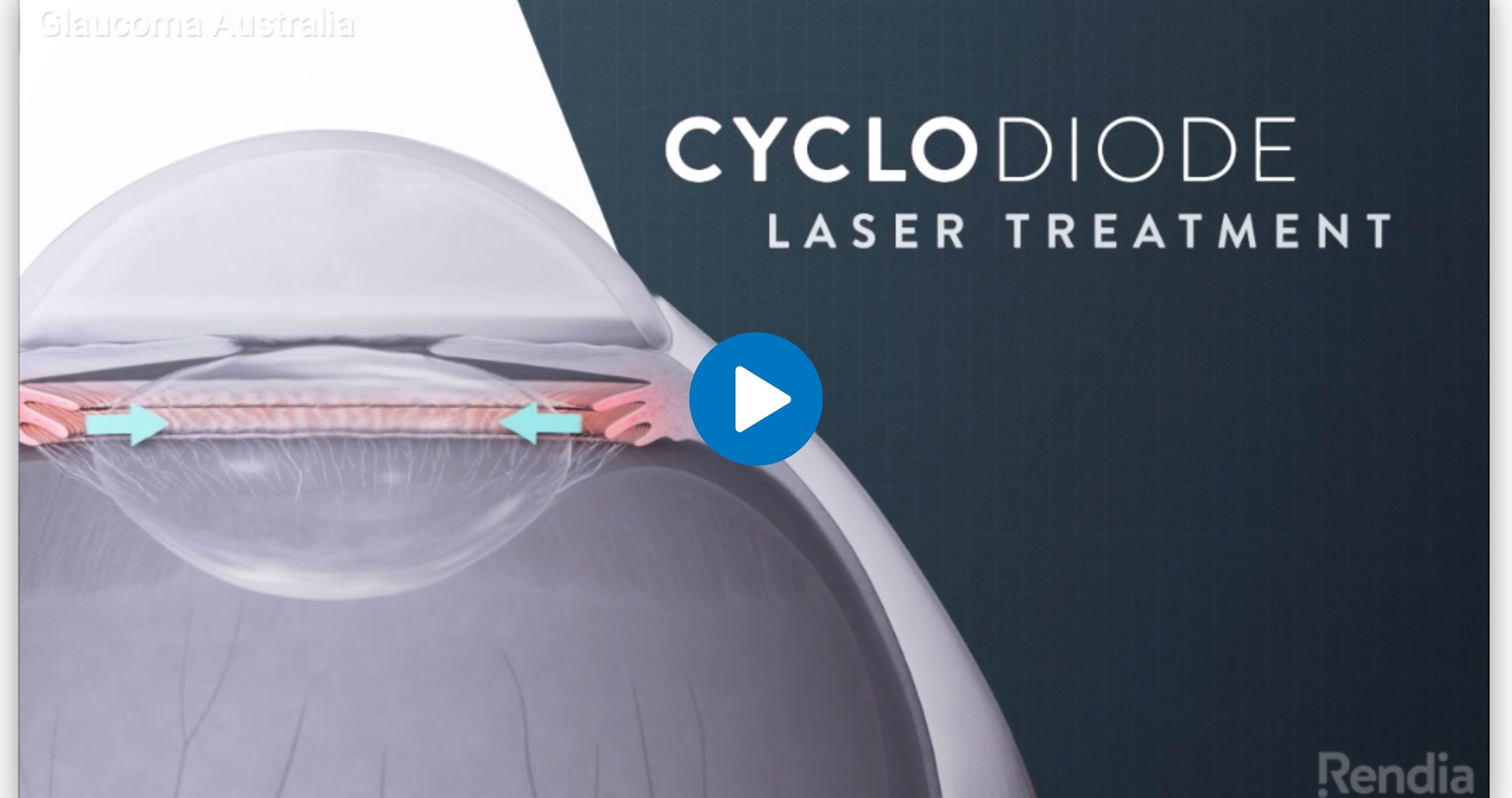
Cyclodiode Laser Treatment
What is it?
Cyclodiode laser treatment causes cyclodestruction, destroying a portion of the ciliary body, a structure in the eye which produces aqueous fluid. This can reduce the amount of fluid produced and therefore reduce pressure inside the eye. The laser energy is directed to the ciliary body via a probe that is held against the wall of the eye.
How does it work?
If the natural (aqueous) fluid that fills the eye cannot drain away properly, it can result in a build-up of pressure within the eye. The high pressure can cause loss of vision and, if very high, pain or discomfort.
Cyclodiode laser treatment works by reducing the amount of aqueous fluid produced and therefore reduce pressure inside the eye.
Who is it suitable for?
This procedure is most commonly used on eyes where other forms of surgery would be difficult or likely to fail. Although the treatment is usually effective, more than one treatment is sometimes required. The aim of glaucoma treatment is to reduce the pressure to a level which is safe for the eye
What are the benefits?
In some patients, cyclodiode laser treatment can have the additional benefit of reducing pain from high pressure.
This procedure DOES NOT improve your vision.
Before the procedure
Please take all medication as normal or as instructed. Your eye specialist (ophthalmologist) will advise you on which of your eye drops to use after the procedure.
During the procedure
This procedure is usually carried out under local anaesthetic. This means that although you will be awake, the eye will be numb so you will not feel any pain. You do not usually have to stay in hospital—the procedure itself lasts approximately 15 minutes and is carried out in the operating theatre.
After the procedure
- You will have an eye shield or pad on your eye which can be removed when you get home. You may notice some blood-stained tears on the eye pad when you remove it. This is normal and there is nothing to worry about
- It is not advisable to drive on the day of the procedure.
- Your sight may be blurred immediately after the operation. This will usually improve after a week or two but can last up to six weeks.
- Your eye will be watery for a short period of time.
- You may have a gritty sensation in the eye for a week or two. Mild pain can be relieved by taking pain killers such as paracetamol.
- Following your operation you should rest and take things easy. You can carry out normal day to day activities.
- You will be given some eye drops to administer regularly. These will usually start on the evening of your surgery once you get home. Your ophthalmologist or discharging nurse will explain the frequency of drops to you.
- If you are on more than one drop wait 5 minutes between each eye drop.
- In addition to your post-operative drops, please continue any regular eye drops in the operated eye until your doctor tells you otherwise. If you take drops in the non-operated eye please continue as normal.
- You will have regular clinic appointments following the surgery to monitor your eye pressures. The first one is normally the next day.
What are the risks?
Any medical treatment involves potential risks. Some of those which apply to this procedure are:
- Pain after the operation.
- Inflammation in the eye.
- In some cases the pressure can be too high or low following treatment.
- High pressure following the procedure may require another treatment session.
- In some cases reduced vision for up to 6 weeks.
- In very rare cases persistently very low pressure can cause permanent loss of vision and alter the cosmetic appearance of the eye.
- Very rarely bleeding or infection.
- Although the risks may sound worrying it is important to remember that cyclodiode laser treatment is generally successful and well tolerated.
Are there any alternatives?
Cyclodiode laser is one of many glaucoma treatments, including eye drops, drainage surgery and other types of laser though it is traditionally reserved for refractory cases of glaucoma. If your Ophthalmologist recommends the use of cyclodiode laser, then alternative treatment options may have proven ineffective, or have been deemed inappropriate. Having an understanding of why cyclodiode is appropriate for you – through discussion with your Ophthalmologist – is recommended.
Newer versions of the cyclodiode laser are available with laser energy delivered via a probe (endoscopic cyclophotocoagulation) or delivered via a series of repetitive shorter pulses of energy (micropulse transscleral cyclophotocoagulation). These have similar efficacy to conventional cyclodiode and may generally be considered for use earlier in the disease process.
Argon Laser Treatment
What is it?
Argon laser is a blue-green spectrum laser which targets pigmented or darker structures in the eye such as the iris.
When, why and how is it done in glaucoma?
Argon laser is used for:
Peripheral Iridoplasty (ALPI):
- WHEN: It is used for selected people with a plateau iris (Zada, Dunn, and White 2018) and occasionally for acute angle closure (Ng, Ang, and Azuara-Blanco 2008)
- WHY: When the edge of the iris is too close to the cornea (clear front window) it risks blocking the drainage of aqueous fluid from the angle of the eye. This can cause the pressure inside the eye (IOP) to increase and worsen glaucoma damage to the optic nerve
- HOW: After local anaesthetic a lens is placed on the surface of the eye to give a clearer view to the doctor and avoid the worry of blinking. The laser is then applied around the edge of the iris to shrink down the thick tissue blocking the drainage angle. There is very minimal discomfort with this procedure, and it takes around 5 minutes per eye to perform.
Argon Laser Trabeculoplasty (ALT):
- WHEN: This procedure has now largely been superseded by selective laser trabeculoplasty (SLT), which is equally effective but leaves much less thermal damage to the eye tissues. Compared to SLT, ALT requires 80-100 times more energy for its pressure lowering effects and generally cannot be repeated. (Kent et al. 2015; Juzych et al. 2004).
- WHY: Indications for this procedure are the same as for SLT. When the IOP is too high and damage to the optic nerve is worsening, your ophthalmologist can improve the drainage of fluid and lower the IOP from the eye using this procedure.
- HOW: After local anaesthetic a lens is placed on the surface of the eye to give a clearer view to the doctor, focus the laser and avoid the worry of blinking. The laser is then applied around the drainage angle. There is very minimal discomfort with this procedure, and it takes around 5 minutes per eye to perform.
References:
Juzych, Mark S., Vikás Chopra, Michael R. Banitt, Bret A. Hughes, Chaesik Kim, Mark T. Goulas, and Dong H. Shin. 2004. “Comparison of Long-Term Outcomes of Selective Laser Trabeculoplasty versus Argon Laser Trabeculoplasty in Open-Angle Glaucoma.” Ophthalmology 111 (10): 1853–59.
Kent, Shefalee S., Cindy M. L. Hutnik, Catherine M. Birt, Karim F. Damji, Paul Harasymowycz, Francie Si, William Hodge, Irene Pan, and Andrew Crichton. 2015. “A Randomized Clinical Trial of Selective Laser Trabeculoplasty versus Argon Laser Trabeculoplasty in Patients with Pseudoexfoliation.” Journal of Glaucoma 24 (5): 344–47.
Ng, W. S., G. S. Ang, and A. Azuara-Blanco. 2008. “Laser Peripheral Iridoplasty for Angle-Closure.” Cochrane Database of Systematic Reviews 2008/07/23 (3): Cd006746.
Zada, Mark, Hamish Dunn, and Andrew White. 2018. “Pilocarpine Test May Predict Success of Argon Laser Peripheral Iridoplasty.” Clinical & Experimental Ophthalmology 46 (9): 1089–91.
