Share
Glaucoma affects an estimated 300,000 Australians but half of them don’t know it. Early detection is considered vital to prevent irreversible vision loss and blindness.
Recognising the limitations of current glaucoma screening techniques, including timeliness and cost effectiveness, researchers have seized on the opportunity to target these shortcomings to design low-cost tests that deliver rapid results.
Not only can their new screening and diagnostic tools detect glaucoma sooner, they could potentially one day form part of national screening programs and alleviate the direct and indirect costs of glaucoma to the Australian healthcare system, projected to hit $784 million by 2025.
This month Insight speaks with three researchers, all non-eye care professionals but experts in their chosen fields, about their work to develop all new imaging devices – and even a blood test – to pick up high-risk individuals before irreversible vision loss sets in.
Fluorescent hyperspectral imaging
Physicist and biomedical engineer Professor Ewa Goldys has collaborated with a team of clinicians, including ophthalmologists, to develop a novel imaging technology for early detection and monitoring.
Goldys is deputy director of the ARC Centre of Excellence for Nanoscale Biophotonics at UNSW that develops novel tools for biomedical diagnostics.
The centre’s research has now extended into the ophthalmic industry where she is working with a team of clinicians – including Clinical Associate Professor Andrew White at Westmead Hospital, Professor Robert Casson, Head of Ophthalmology and Visual Sciences at the University of Adelaide, and UNSW Scientia Fellow Dr Nicole Carnt – to develop a bespoke camera that will help ophthalmologists measure the oxidative stress of cells and tissues in the retina.
“Ophthalmology is an area of clinical medicine producing extensive images of the eye – even mobile phone cameras today are equipped with amazing image capability. At the same time, science is developing better technical analysis of images. We’re building on that particular junction in time to provide deeper image analysis,” Goldys says.
In 2021, she was awarded Glaucoma Australia (GA)’s ‘Quinlivan’ Research Grant to support her research in fluorescent imaging technology to detect glaucoma.
Goldys’ imaging innovation obtains information about the health status of the retina and the optic nerve, providing the opportunity for early disease detection and the ability to commence treatment before irreversible blindness sets in.
“Neurons die, which is irreversible. We need to know when do neurons start to be in trouble?,” she says. “When there is progressive loss of vision, it is clearly too late. We need to address vision loss earlier. This represents an unmet need.”
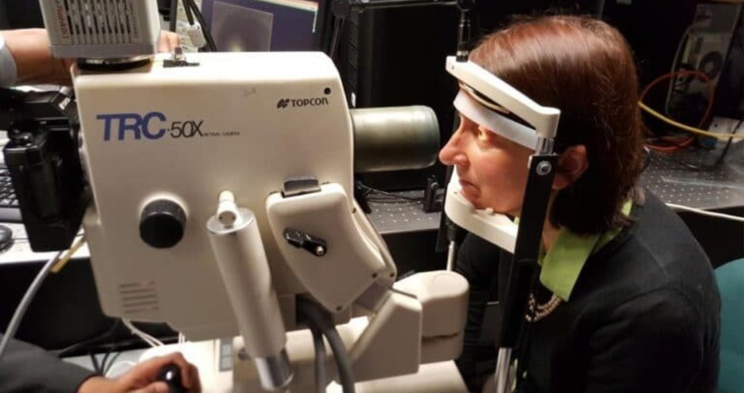
Refurbishing current technology that is user-friendly and non-invasive, Goldys and her team are photographing the eye, then splitting the image into different colour segments.
“Our lab is full of bespoke imaging systems. We are currently refurbishing and re-engineering a fundus camera for trials with a small Australian company called Quantitative Pty Ltd,” she says.
“It’s like shining sunlight through a prism. Once separated into individual colours, light provides nuanced information. We’re interpreting colours and shapes of cells, by a method that links colour to specific molecules, which are central to metabolism.
“Cells need energy to survive. At a cellular level, neurodegeneration is a symptom of cells losing energy supply.”
As part of their pre-clinical research, Goldys and her team are currently testing metabolism in cells that are affected, and are interfering with cells by exposing them to oxidative stress.
“We have proven oxidative stress is inducing colour change in cells. Our study on this was published in peer-reviewed journal Redox Biology, which is not an ophthalmology journal,” Goldys says, adding she is aware of the debate in ophthalmology about countering oxidative stress by supplementing with antioxidants.
Their proof-of-concept study demonstrated that an imbalance of unstable molecular species called ‘free radicals’ will change the colour of cells – and a new imaging technique could allow clinicians to detect and decode this colour without needing to take samples from the body.
“In our study of cell cultures and tissues in the lab, we found that colour is like a thermometer for oxidative stress,” Goldys says.
With the support of the ‘Quinlivan’ Research Grant for the next two years, Goldys hopes to have finished pre-clinical trials within one year.
Related Articles
Part 2: Genetic testing for degenerative eye disease
Part 3: Computerised glaucoma screening test
Published with permission from www.insightnews.com.au

Researchers have used mice with nine different genetic backgrounds to identify factors influencing eye ageing, paving the way for eye-based diagnostics for neurodegenerative diseases.

Researchers have found that a vitamin supplement that improves metabolism in the eye appears to slow down damage to the optic nerve in glaucoma.
Long-term variability in blood pressure may be associated with visual field (VF) progression in glaucoma patients, suggesting new considerations for managing this sight-threatening condition...