Share
With no community wide screening program in place, Australia and New Zealand rely on opportunistic screening to increase detection and reduce progression of glaucoma. As the incidence of this ‘sneak thief of sight’ continues to escalate, it’s a job that requires the commitment of optometrists and ophthalmologists, supported by increasingly sophisticated technology
![]()
Between 2.7 to 3.1 per cent of the Australian population over 50 years of age have glaucoma or are strong suspects.1 Additionally, 5.7 per cent have ocular hypertension without glaucoma. Both these numbers are predicted to rise with the ageing Australian population.1 Over 50 per cent of glaucoma in Australia is likely to be undiagnosed.2
Current workforce calculations suggests that neither optometrists nor ophthalmologists alone are capable of screening, or even reviewing these patients at the recommended biannual interval.3 This makes a collaborative approach to glaucoma diagnosis and care essential – not only because of the necessity arising from the volume of patients to be screened and reviewed; but also because we need to achieve the benefits of efficient patient care, and timely diagnosis via opportunistic screening.
Fortunately, in Australia, we have a large and widely spread optometry workforce that is willing to work with ophthalmology to detect and manage glaucoma. There are now over 5,000 optometrists, with more than half endorsed to prescribe glaucoma medication. Their services are covered by Medicare and most bulk bill examinations. While there is still unmet demand for regional services, optometrists are found in most parts of the country, and almost all practices continue to accept new patients with appointments available throughout the week.
At present there is no community-wide screening program for glaucoma in Australia. Instead opportunistic screening by optometrists and ophthalmologists helps reduce the sojourn time during which a patient with glaucoma can be picked up and treated before their disease becomes symptomatic.
Diagnosis and Monitoring
Glaucoma is diagnosed and monitored with a combination of functional and structural investigations, based on clinical history and examination.4 The current technologies for investigating optic nerve and retinal ganglion cell structure and visual function all have high false positive rates, and therefore none should be used in isolation. Clinical acumen remains important to correctly diagnose glaucoma, for both ophthalmologists and optometrists.3
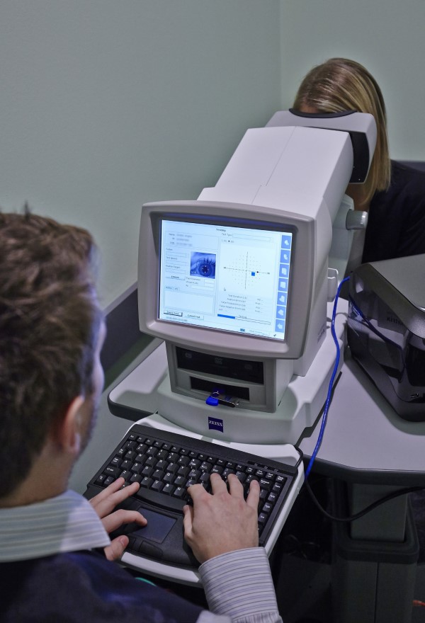
The standard optometry practice is equipped to detect glaucoma – the basic tools for detection are an ability to assess the optic nerve, measure the pressures and analyse the visual fields. As the majority of patients examined by optometrists do not have glaucoma, practices are usually designed to screen for glaucoma risks. Tools that are most suitable for glaucoma screening have good patient acceptance, are economical and have reasonable sensitivity. These tools may be different to those considered gold standard for a glaucoma work-up, however, when used as a battery of tests, they provide a good basis for identifying those at risk of glaucoma so that additional investigation can be provided to more accurately determine whether the condition is present.
Intraocular Pressure
Intraocular pressures (IOP) are a standard part of the optometry eye examination. Many patients will have their pressures screened by non-contact tonometry (NCT). This has excellent patient acceptance and is economical due to its multifunctionality and speed. However, NCT has significantly higher variability and is less reliable outside the physiological range of IOP, which is why most optometrists will also have an applanation tonometer for use on indication.
Central Corneal Thickness
Central corneal thickness (CCT) should be checked in all those with risk factors for glaucoma. IOP should be interpreted in the context of the patient’s CCT, and CCT may be valuable to personalise target pressures for our patients. For example, in discontinuing therapy in a patient with ocular hypertension treatment and low risk of glaucomatous damage, who is found to have thick CCT; or escalating management in a patient with thin CCT.5
Visual Fields
The visual field is the best functional measure of our patients’ vision despite variability in measurements. Because of this, computerised automated perimetry is the cornerstone of interpreting glaucoma, as it measures direct visual impact rather than indirect measures such as IOP and optic nerve changes5
Frequency Doubling Technology relies on flicker detection and is therefore not directly comparable to other visual fields. It is more sensitive for the detection of glaucoma, but less reliable in monitoring than standard automated perimetry.
Imaging Technologies
While experienced examination and photography of the optic disc is effective in detecting glaucomatous damage, the 2016 World Glaucoma Association consensus statement suggested that, “Imaging technologies, including optical coherence tomography (OCT), confocal scanning laser ophthalmoscopy (such as Heidelberg Retina Tomograph HRT) and scanning laser polarimetry (such as GDx) have afforded a more objective and quantitative approach to detect and monitor glaucoma.”
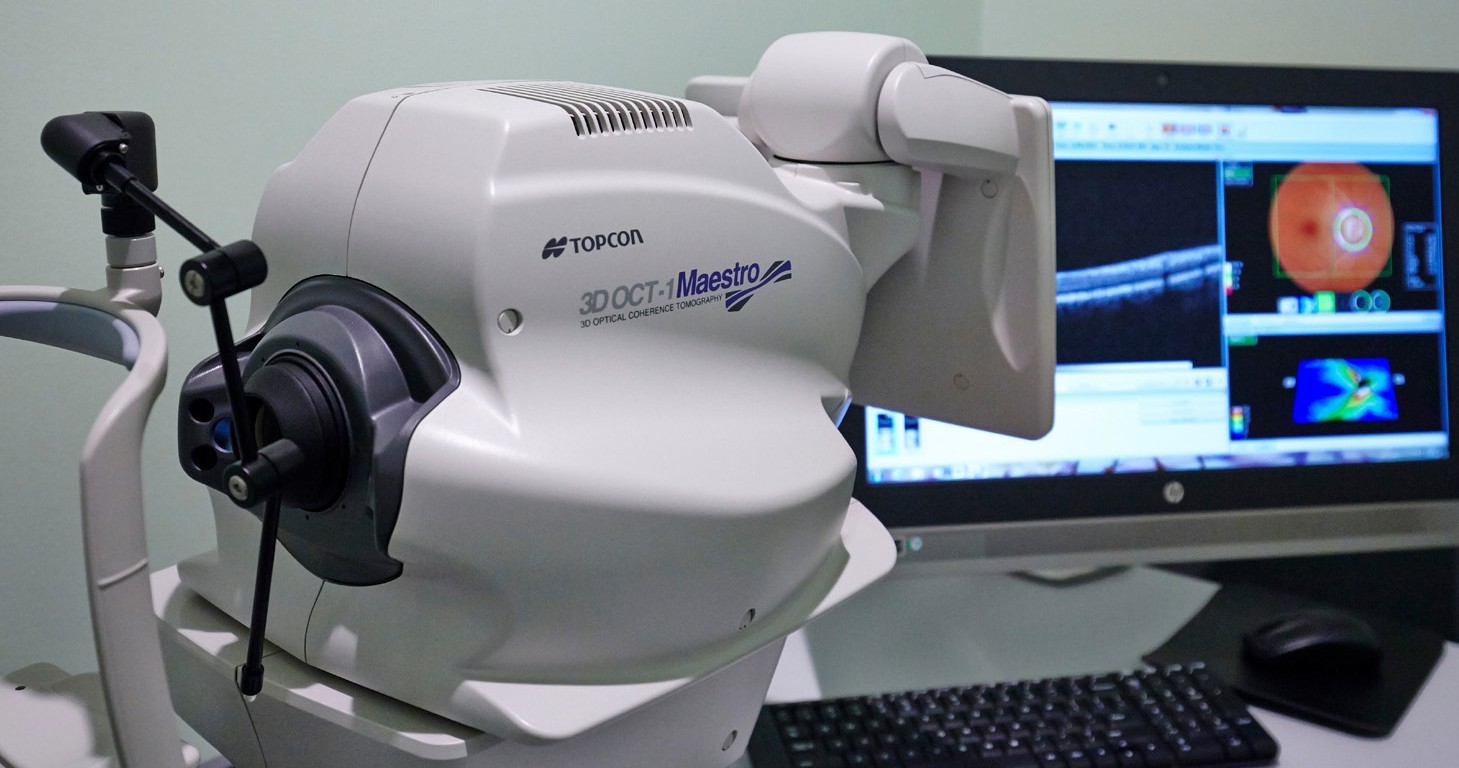
The sensitivity and specificity of OCT, GDx and HRT are approximately 80 per cent with an inter-device agreement between 25-40 per cent.4 The 2016 WGA consensus on glaucoma diagnosis states, “OCT measurement of retinal nerve fibre layer (RNFL) thickness may be the best among the currently available digital imaging instruments for detecting and tracking optic nerve damage in glaucoma”.
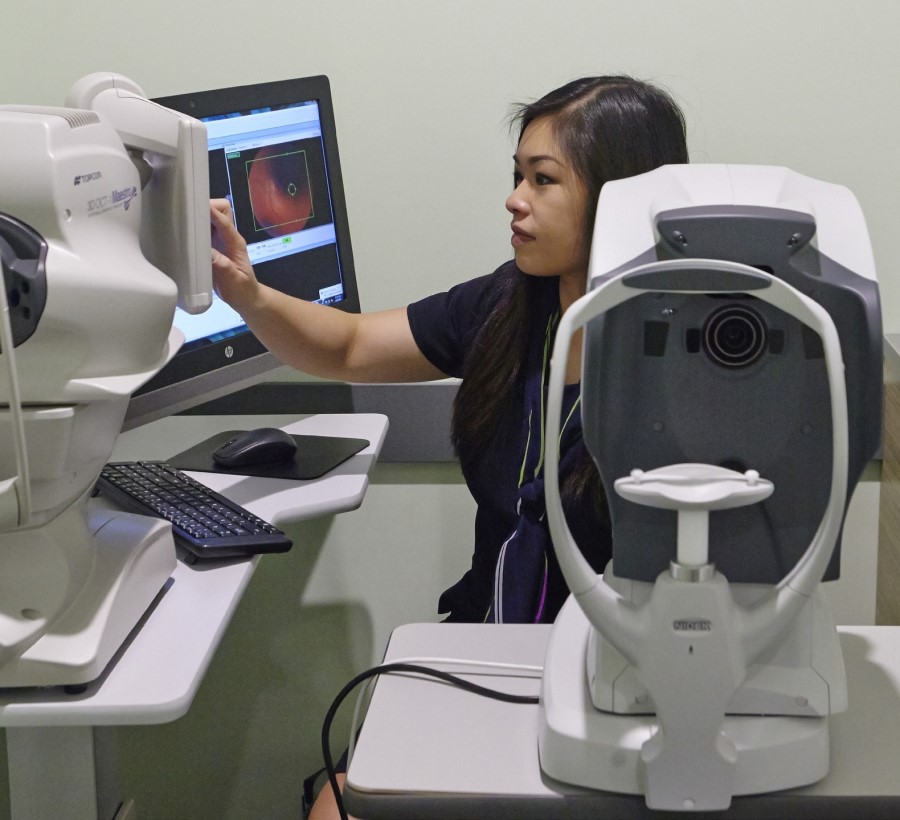
Retinal photography is however, a more widely available tool for optic nerve assessment. Although this may not have the benefit of the same level of analysis and ability to isolate structures as OCT, serial analysis of retinal photos over time has proven to be a sensitive technique to detect glaucoma and its progression.6 Furthermore, advances in machine learning have already demonstrated detection rates comparable with specialists for diabetic retinopathy using retinal photographs, with these same researchers also working on developments for glaucoma.
A barrier to combining the strengths of ophthalmology and optometry to defeat glaucoma can be a failure to effectively share patient information
Collaborative Care
Optometrists with therapeutically- endorsed training may manage glaucoma suspects and glaucoma patients identified as stable in collaboration with ophthalmology colleagues.3
A barrier to combining the strengths of ophthalmology and optometry to defeat glaucoma can be a failure to effectively share patient information. This can result in unnecessary repeating of tests, delays in diagnosis, loss to referral, loss to follow- up and additional costs. The use of an eReferral platform, such as Oculo, is one possible solution to facilitate safe and efficient sharing of patient information, leading to better care and patient outcomes.
Duplication of services is both a frustration for patients and unnecessary cost to the healthcare system. A framework of responsibilities for each carer is a major step in overcoming this challenge. This is ideally a local arrangement between professionals based on their specific circumstances. The ideal starting point for such agreements is evidence based guidelines, such as those of the NHMRC and the RANZCO glaucoma referral pathways. These are also the foundation blocks of greater inter-professional consistency.
There is a great opportunity across Australia and New Zealand for ophthalmology and optometry to work together to improve glaucoma detection and treatment. Forecasts suggest neither workforce can care for this population alone.7 Both professionals have tools appropriate for the task and specific to their mode of service delivery. With the wider distribution of optometry, and the deeper specialisation of ophthalmology, co-management appears to be an effective way to tackle this condition. Optometry is best positioned for detection and management of chronic stable glaucoma, which frees more of ophthalmology’s resources for complicated and surgical cases. Placing the patient first in the shared care of glaucoma will often mean local professionals must adopt systems and protocol that make the best use of their specific skills and resources.
There is a great opportunity across Australia and New Zealand for ophthalmology and optometry to work together to improve glaucoma detection...
References
-
Rochtchina, E. and P. Mitchell, Projected number of Australians with glaucoma in 2000 and 2030. Clin Exp Ophthalmol, 2000. 28(3): p. 146-8.
-
Mitchell, P., et al., Prevalence of open-angle glaucoma in Australia. The Blue Mountains Eye Study. Ophthalmology, 1996. 103(10): p. 1661-9.
-
White, A., et al., Guidelines for the collaborative care of glaucoma patients and suspects by ophthalmologists and optometrists in Australia. Clin Exp Ophthalmol, 2014. 42(2): p. 107-17.
-
Society, A.P.G., Asia Pacific Glaucoma Guidelines. 2016, Kugler Publications: Amsterdam, The Netherlands.
-
Shaarawy, T., et al., eds. Glaucoma: Medical diagnosis & therapy. Second ed. Vol. 1. 2015, Elsevier.
-
Heijl A, Bengtsson B, Diagnosis of early glaucoma with flicker comparisons of serial disc photographs. Invest Ophthalmol Vis Sci 1989;30 (11) 2376- 2384
-
White A, Goldberg I, Guidelines for the collaborative care of glaucoma patients and suspects by ophthalmologists and optometrists in Australia. Clin Exp Ophthalmol. 2014 Mar;42(2):107-17


Mike Amor and Estelle Powell have never met, yet their stories share a common thread that should give every eye care practitioner pause for thought. Both experienced critical moments where the system – and the people within it – either saved or compromised their vision.
mivision congratulates Emeritus Professor Leo Carney, Professor Bill Huxley Morgan, and Professor Konrad Pesudovs who were awarded in the King’s Birthday 2026 Honours List.
A United States ophthalmologist has unveiled a novel surgical technology that allows eye surgeons to measure and respond to critical fluid dynamics inside the eye in real time - an advance that may significantly improve precision and outcomes in glaucoma and other ophthalmic procedures.