Share
Cutting-edge technology offers non-invasive, real-time measurement of retinal metabolism and reveals subtle changes in cellular energy metabolism.
Glaucoma detection and monitoring have reached new horizons with the pioneering application of hyperspectral imaging of cellular autofluorescence. This innovative technology allows scientists to examine the natural glow, called autofluorescence, emitted by cells in the eye to identify early signs of glaucoma. Hyperspectral imaging can reveal detailed information about the different colours emitted by cells, and therefore, allows researchers to spot subtle changes in how cells produce and use energy, offering the potential to detect glaucoma before it causes irreversible vision loss.
Glaucoma, a leading cause of blindness worldwide for people over the age of 50, damages the optical nerve. Approximately 300,000 Australians and more than 70 million people globally are affected by glaucoma, but alarmingly, half of them remain unaware of their condition, mistakenly believing that they have healthy eyes. Research has highlighted the significance of energy insufficiency accompanying neurodegeneration and mitochondrial dysfunction as a key driver of glaucoma pathogenesis.
“By focusing on this critical aspect, our solution opens new avenues for early intervention. The core of our breakthrough lies in our ability to utilise hyperspectral imaging to map spectral signatures of energy metabolites in a retinal cell culture and tissue models. This enables us to investigate alterations induced by known stressors, shedding light on the early stages of glaucoma development.” says lead author of the research and optometrist Abhilash Goud Marupally from UNSW.
"Our goal is to use the unique capabilities of hyperspectral imaging to detect glaucoma early, when treatment options are most effective. By understanding how cells produce energy and monitoring these changes over time, we can make a real difference in preserving vision." senior researcher on the project at UNSW and primary investigator, Scientia Professor Ewa Goldys, noted.
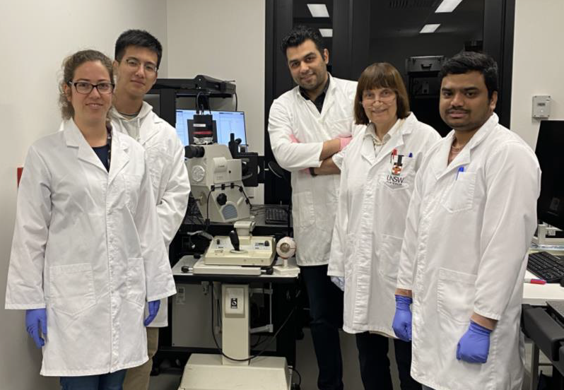
In a remarkable collaborative effort funded by Glaucoma Australia’s ‘Quinlivan’ Research Grant, researchers at UNSW joined forces with esteemed partners and experts in the field of glaucoma research. The project has brought together the clinical expertise, knowledge, and invaluable advice of multiple collaborators, including glaucoma specialists Associate Professor Andrew White, Head of Department of Ophthalmology at Westmead Hospital, Sydney; Professor Robert Casson who leads the Department of Discipline of Ophthalmology & Visual Science at the University of Adelaide; and Scientia Associate Professor Nicole Carnt, Deputy Director of Research at the School of Optometry and Vision Science, UNSW.
"Our partnership allows us to include diverse perspectives and expertise, resulting in a comprehensive approach to studying glaucoma. Through our collective efforts, we aim to translate scientific discoveries into meaningful clinical applications. The collective knowledge and experience of our collaborative team have been instrumental in guiding this research. Our aim is to bridge the gap between research and clinical practice, with the ultimate goal of improving the lives of individuals with glaucoma.“ Scientia Professor Ewa Goldys states.
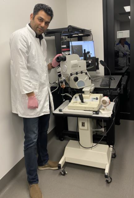
The team is also developing a modified fundus camera system that can capture images of the back of the eye. This affordable modification of an off-the-shelf device allows for the integration of autofluorescence hyperspectral imaging capabilities, making it a cost-effective solution for real-time measurement of retinal metabolism translatable to clinical practice.
"Our goal was not only to advance glaucoma detection technology but also to ensure its practicality and ease of implementation in real-world clinical settings. By remodelling the Fundus camera, we have created a solution that can seamlessly integrate into existing diagnostic workflows, providing ophthalmologists and optometrists with valuable insights for glaucoma diagnosis and monitoring.", Abhilash Goud Marupally says.
Professor Ewa Goldys shared her excitement about the potential impact of this research, stating, "The development of the modified fundus camera represents a significant advancement in glaucoma monitoring. By utilising a readily available device and integrating hyperspectral imaging technology, we can use the benefits of real-time retinal metabolism measurement for screening purposes of a broad population. This technology will allow clinicians to track metabolic changes in real-time and offer personalised treatments to their patients. Our cost-effective implementation of hyperspectral autofluorescence imaging into regular fundus camera systems holds tremendous promise for improving patient outcomes and transforming glaucoma management leading to immense socio-economic benefits."
Dr. Abbas Habibalahi, the researcher managing the translatable research project at UNSW, expressed his enthusiasm, saying, "The application of hyperspectral imaging in glaucoma research is a game-changer. It has the potential to transform the way we diagnose and manage this sight-threatening condition. By utilising this cutting-edge technology, we can make significant strides in preserving vision and improving the quality of life for individuals with glaucoma and potentially also use this technology to investigate and diagnose other diseases of the eye."
Professor Goldys notes that, in the near term, the team, together with their esteemed collaborators, will continue to work on optimising the fundus camera system and extend their investigations to tissue samples to collect clinically relevant data and validate the potential of hyperspectral autofluorescence imaging as a diagnostic tool. By conducting extensive clinical studies and collaborating with renowned experts, they want to ensure that this innovative technology can be seamlessly adopted by ophthalmologists and glaucoma specialists worldwide. Moreover, the potential applications of this system extend beyond glaucoma. With further research and development, this technology could potentially be utilised for the diagnosis and monitoring of other ocular conditions, such as eye cancer.

Researchers have used mice with nine different genetic backgrounds to identify factors influencing eye ageing, paving the way for eye-based diagnostics for neurodegenerative diseases.

Researchers have found that a vitamin supplement that improves metabolism in the eye appears to slow down damage to the optic nerve in glaucoma.
Long-term variability in blood pressure may be associated with visual field (VF) progression in glaucoma patients, suggesting new considerations for managing this sight-threatening condition...