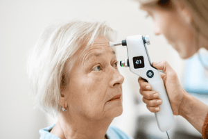
Glaucoma occurring with Sturge-Weber Syndrome (SWS) is one of the most challenging sorts of glaucoma for the eye specialist to treat.
SWS is a congenital condition which can have ocular, neurological and skin manifestations. It is present at birth, but it is not a hereditary or genetic condition. It often involves the skin of the scalp and face, around the eye, on one side. There are masses of abnormal dilated veins which on the skin are called a naevus flammeus or port-wine stain - a dark crimson discolouration which does not increase in extent after birth, but may become thicker and darker. It is not a tumour in the sense of an abnormal growth of tissue, or a cancer.
The dilated blood vessels also exist inside the skull and can involve brain tissue, sometimes causing epilepsy, hemiplegia or cognitive deficit. With regard to the eyes, the dilated blood vessels can occur both inside and outside the eyeball. Outside, the eyelids and/or the conjunctiva can be affected. If the conjunctiva is affected the eye will be “blood-shot”. Inside, the vascular layer of the eyeball (the choroid- a choroidal haemangioma) can be involved and if it is, glaucoma is more likely, and other complications can occur.
Glaucoma in SWS occurs because of the abnormal blood vessels in and around the eye. The increased blood flow through the choroid tends to make more pressure in the eye, and high vascular pressure outside the eye impairs normal drainage of fluid from the eye, also pushing the pressure up. Ideally, glaucoma in SWS would best be treated by removing the abnormal vessels but unfortunately this is not technically possible. Even if the eyelid skin is treated with laser, to make the haemangioma less apparent, it does not alter the blood flow around the eye and does not alter the glaucoma.
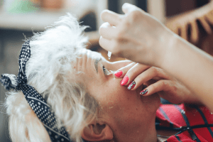
If glaucoma occurs, the first line of treatment is with drops, such as timolol or latanoprost, but pressure control is often difficult and surgery may be necessary. Such surgery could be a filtering operation (trabeculectomy) which creates a filtered hole for fluid to drain from the inside of the eyeball to the tissues just under the surface layer of the eyeball (conjunctiva).
Children generally have a very active healing response, and the filtered hole is prone to healing over so certain drugs (mitomycin-c or 5-FU) may be used at the time of surgery to suppress this healing response. If a filtering operation is insufficient to control the intraocular pressure, a tube implant (eg Molteno or Baerveldt) may be placed in the eye. The glaucoma which can occur in SWS is not treatable with laser (ALT).
Children with SWS may lose vision due to uncontrolled glaucoma, and also due to the leakage of fluid from a choroidal haemangioma under the retina. If this leakage occurs, sometimes it can be successfully treated with retinal laser therapy. Eyeballs affected by glaucoma in childhood, are often larger than normal, and are prone to become short-sighted, so glasses are often necessary.
The management of glaucoma in SWS has improved immensely over the past decade, but still remains a challenge. Fortunately the condition only affects one eye, and children affected by SWS are not visually handicapped.